Healthy vs. Diseased Kidney
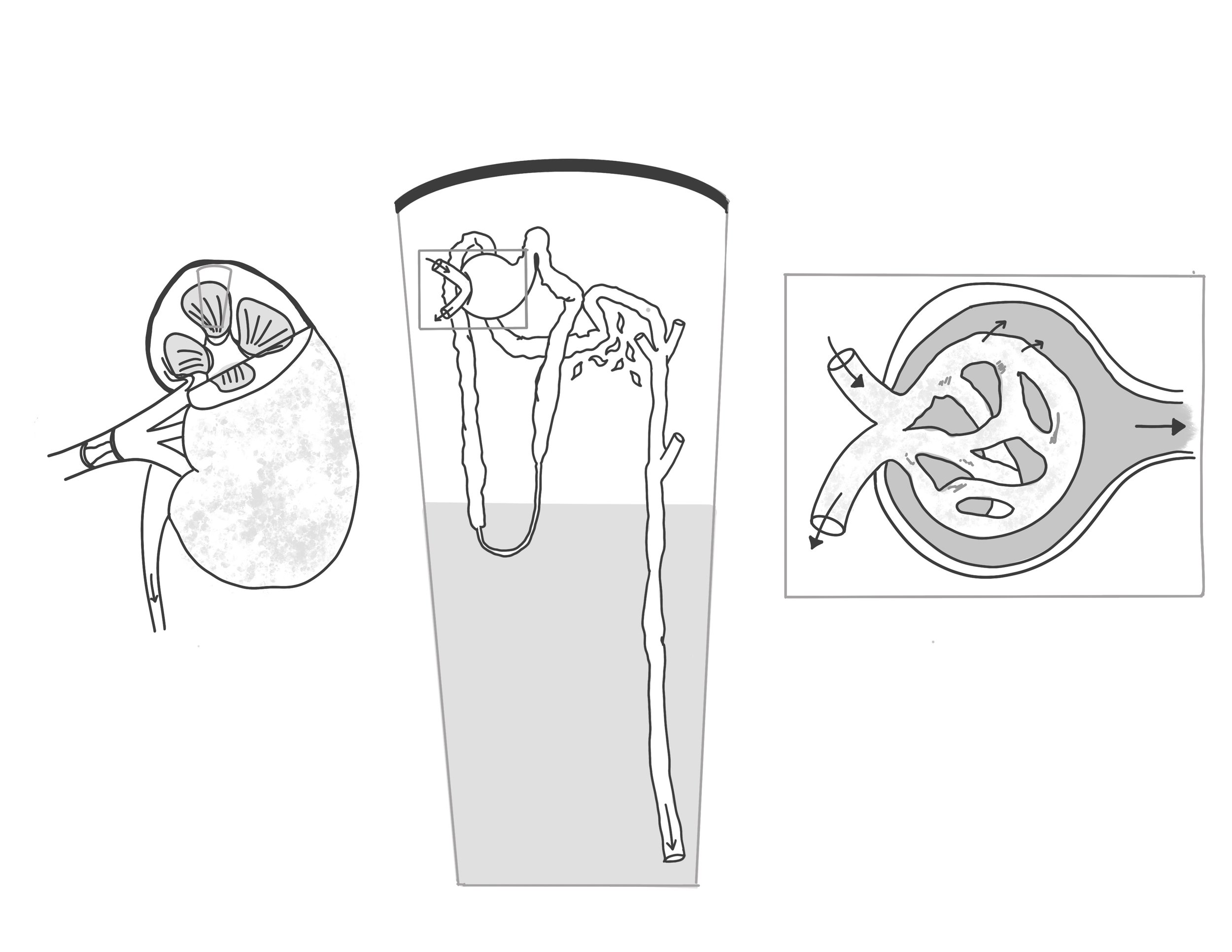
The goal of these illustrations was to show a healthy vs. diseased kidney at the gross level, nephron level, and glomerulus level for digital presentation. Labels were omitted in the final deliverables, allowing flexibility for the client when incorporating the illustrations into his presentations.
Client: Rohit Singh MD/PhD Student
Media: Procreate, Adobe illustrator
Format: Digital (eg. in a PowerPoint slide)
Audience: Educated lay audience
Date: April 2021
Healthy Kidney
Diseased Kidney
Process work
Research
I reviewed references provided by the client and anatomical textbooks to understand the general changes that occur in diseased kidneys in comparison to healthy kidneys. This included changes in size, texture, blood flow, atrophy, scarring, etc.
Layout + Sketches
Revised Sketches

Final Rendering
The illustrations were first rendered in grayscale to establish the dark and light values. Then, they were colorized by gradient mapping in Procreate and brought into Adobe Illustrator for the addition of arrows and shape outlines.


References
Agur, A. M. R. (2022). Grant’s Atlas of Anatomy (Fourteenth, International ed.). LIPPINCOTT.
Chronic kidney disease - Symptoms and causes. (2021, September 3). Mayo Clinic. https://www.mayoclinic.org/diseases-conditions/chronic-kidney-disease/symptoms-causes/syc-20354521
Gilroy, A. M., MacPherson, B. R., Ross, L. M., Schuenke, M., Schulte, E., & Schumacher, U. (2012). Atlas of Anatomy (2nd ed.). Thieme.
Md, F. N. H. (2022). Netter Atlas of Human Anatomy: Classic Regional Approach: paperback + eBook (Netter Basic Science) (8th ed.). Elsevier.